Products in stock in the United States can be delivered within 2 days.
 +1-2404726069 (U.S.) +86-027-65528241 (Outside U.S.)
  overseas@yeasen.com
 0
0SuperSignal MaxiSignal Maximum Sensitivity Substrate

Product Description
SuperSignal MaxiSignal Maximum Sensitivity Substrate is designed to detect antibodies and associated antigens directly or indirectly labeled with horseradish peroxidase (HRP). The principle is that, proteins or nucleic acids were transferred to the imprinted membrane after electrophoresis, and the target proteins on the membrane were bound by primary antibody and secondary antibody labeled with HRP, or the nucleic acids on the membrane were bound directly or indirectly by probes labeled with HRP. After washing the membrane, the ECL working solution prepared by the product was used to incubate the membrane at room temperature for several minutes. The imprinted membrane was wrapped with plastic wrap and fixed to the X-ray exposure Cassette. Then the X-ray film is pressed on the membrane in a darkroom and exposed for several seconds to several hours. After development and fixing, protein or nucleic acid bands can be clearly displayed on the X-ray film.
Product Features
1. High sensitivity and high signal-to-noise ratio, it can detect low fick-grade antigens.
2. Long duration of luminescence, fluorescence can make X-ray film sensitive for more than 8 hours, especially suitable for trace protein or nucleic acid detection.
3. Higher dilution ratio of antibody can be used to greatly save antibody.
Dilution ratio of primary antibody (liquid storage concentration 1mg/mL) : 1:1000-1:100000.
Dilution ratio of secondary antibody (liquid storage concentration 1mg/mL) : 1:100000-1:500000.
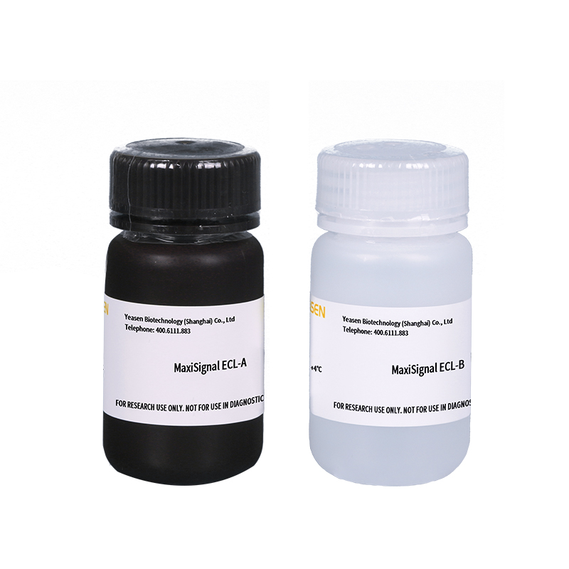
Product components
|
Component |
36224ES10 (10 mL) |
36224ES60 (100 mL) |
36224ES70 (200 mL) |
36224ES76 (500 mL) |
|
|
36224-A |
MaxiSignal ECL - Reagent A |
5 mL |
50 mL |
100 mL |
250 mL |
|
36224-B |
MaxiSignal ECL - Reagent B |
5 mL |
50 mL |
100 mL |
250 mL |
Shipping and Storage
The products are shipped with ambient temperature and can be stored at 4℃ for one year.
[Notes]: Reagent A (36224-A) should be stored away from light!
Instructions
Ideal Western blot results require optimization of all experimental links involved, such as sample loading amount, gel type, membrane transfer method, membrane type, blocking reagent, concentration of primary antibody, concentration of secondary antibody and corresponding incubation time, etc. In addition, sufficient amount of incubation reagents should be provided for each link to avoid membrane drying.
1. Routine electrophoresis, transfer membrane, antibody labeled with HRP or nucleic acid probe labeled with HRP incubation and washing membrane.
[Notes]: ECL luminescent solution is the color substrate of HRP, so the detection system must be based on HRP enzyme-labeled antibody or nucleic acid probe.
2. At the same time of washing the film for the last time, the luminescent working solution was prepared fresh: take the same volume of Reagent A and B, mix well, and place at room temperature for using later.
[Notes]: Take Reagent A and Reagent B must use different tips.
3. Use flat tweezers to take out the membrane, put it on filter paper to drain the lotion, do not make the membrane completely dry. Completely immerse the membrane in luminescent working solution (100-200 μL luminescent working solution/cm2 membrane),
sufficient contact with luminescent working fluid. Incubate at room temperature for 3-5 mins and prepare for tablet exposure immediately.
[Notes]: long incubation time will not increase sensitivity, and sometimes will lead to abnormal exposure bands. The nature of the luminescence process is an enzymatic reaction that uses little luminescence to work, the adverse reaction of the liquid will also lead to uneven exposure of the strip on the film and significantly reduce the sensitivity. In order to achieve the purpose of saving the film can be cut small, but do not reduce the luminous liquid use ratio.
4. Pick up the membrane with tweezers and lay it on filter paper to drain the luminescent working fluid. But do not wash off the luminescent fluid.
5. Lay a piece of plastic wrap larger than the membrane on the inner surface of the X-ray film obscura. Attach the imprinted membrane to the plastic wrap, fold the plastic wrap completely around the imprinted membrane, and remove the gas bubble and fold, can cut off the edge of excess plastic wrap. Use filter paper to suck up excess luminous working fluid. Fix the plastic wrap covering the imprinted membrane in the obscura with tape, with the protein band facing upwards.
6. In the darkroom, take a piece of X-ray film and place it on the wrapped membrane. Press the film and expose it for different times, such as a few seconds to a few minutes. Then the film was developed and fixed.
[Notes]: The exposure time should be adjusted according to the exposure intensity. If the background is too high, use two X-ray films at the same time.
Cautions
1. For your safety and health, please wear lab coats and disposable gloves for operation.
2. Steps 1 to 5 can be operated under fluorescent lamps. However, the sensitivity of luminescent fluid exposed to strong light for a long time may be slightly reduced, which can be avoided by moving to the obscura. Wearing gloves will do avoid leaving fingerprints on the membrane and keep it clean.
3. Prolonged exposure or excessive protein will deepen the background and make the change of band strength lose linear relation. Underexposure will blur the bands.
4. After incubation with the luminescent working solution for about 3 minutes, the bands on the film were luminous. The strong bands were visible in the obscura, while the low abundance bands were weak or even invisible to the naked eye, but the X-ray film can be exposed. The luminous time of bands cannot be judged simply by naked eye observation. The invisible fluorescence lasts for hours and makes the X-ray film feel Light, so weak bands can be exposed for 1~10 hours. If the band is not good after exposure, the membrane can be washed with a washing buffer, the secondary antibody can be incubated again, and ECL can be used again to exposure.
5. As hypersensitive luminescence solution is extremely sensitive; it is highly recommended that the initial concentration of most imported antibodies be 1:1000~1:100000 for primary antibody (liquid storage concentration 1mg/mL) and 1:50000~1:500000 for secondary antibody (liquid storage concentration 1mg/mL). Higher dilution ratio of antibody will result in a high background or no bands, resulting in failure.
6. Some plastic wrap may quench the fluorescence when wrapping the imprinted membrane. High quality plastic wrap should be selected.
7. Avoid placing multiple membranes in the same washing membrane box, as mutual adsorption or friction may cause a deep background.
8. The position and size of the bands on the film can be accurately determined using visible pre-stained protein markers and fluorescent-autoradiography exposure tags.
9. Use a biotin-avidin system and avoid using milk closure, which may cause the background to be too high.
10. Metal oxide particles may cause granular spots on the membrane. Avoid using rusty scissors and tweezers. Use plastic flat tweezers.
11. Sodium azide (NaN3) can inhibit HRP activity, if the recovery of HRP labeled probe or antibody should avoid using NaN3, if necessary, not more than 0.01%.
12. This product has no special toxicity, according to common chemical treatment.
13. For research use only!

[1] Zhang Y, Liu F, Feng Y, et al. CircRNA circ_0006156 inhibits the metastasis of prostate cancer by blocking the ubiquitination of S100A9 [published online ahead of print, 2022 Jun 27]. Cancer Gene Ther. 2022;10.1038/s41417-022-00492-z. doi:10.1038/s41417-022-00492-z(IF:5.987)
[2] Yu Y, Fang L. CircRPAP2 regulates the alternative splicing of PTK2 by binding to SRSF1 in breast cancer. Cell Death Discov. 2022;8(1):152. Published 2022 Apr 2. doi:10.1038/s41420-022-00965-y(IF:5.241)
[3] Cao Y, Sun M, Wang J, Hu X, He W, Su J. Phenotypic and genetic characterisation of an emerging reovirus from Pekin ducks in China. Sci Rep. 2019;9(1):7784. Published 2019 May 23. doi:10.1038/s41598-019-44178-3(IF:4.011)
[4] Sun L, Song F, Liu H, et al. The novel mutation P36R in LRP5L contributes to congenital membranous cataract via inhibition of laminin γ1 and c-MAF. Graefes Arch Clin Exp Ophthalmol. 2020;258(12):2737-2751. doi:10.1007/s00417-020-04846-x(IF:2.396)
Catalog No.:*
Name*
phone Number:*
Lot:*
Email*
Country:*
Company/Institute:*
